


Injury Information
Questions on Your Injury?Where Does It Hurt?
Roll over the image below to learn more about injuries to different parts of the body or click the links for the body part you would like to learn more about.

-
Neck
-
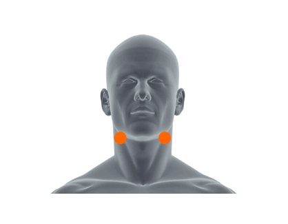
The spinal column is composed of a cervical vertebrae, thoracic vertebrae, lumbar vertebrae, and sacral bones. In between each vertebrae lies an intervertebral disc which cushions the spine and acts as a shock absorber with movement.
Herniated Disc:
When a cervical disc becomes herniated, the disc can press on nerves and cause pain, numbness and tingling. A disc can be damaged by a fall or accident, repeated straining of the neck and lifting of heavy objects. However, disc injuries can also occur over time, without any specific trauma.
Symptoms:
» Aching neck pain
» Numbness, tingling, pain, weakness in one or both arms
» Inability to straighten neck without extreme pain
Neck Strain & Spasms
When a strain occurs in muscles, it is usually accompanied by the tearing or stretching of the muscle fibers. Neck strains can result from whiplash or lifting heavy objects. A spasm can occur when the neck muscles contract involuntarily either as the result of an injury, overuse, poor posture or stress.
Symptoms:
» Pain and tenderness in your neck
» Pain when moving your head
» Tension headaches
» Hard, tight and painful muscles
-
-
Back
-
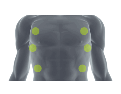
The spinal column is composed of cervical vertebrae, thoracic vertebrae, lumbar vertebrae and sacral bones. In between each vertebrae lies an intervertebral disc which cushions the spine and acts as a shock absorber with movement.
Herniated Disc:
When a disc becomes herniated, this disc can press on nerves and cause pain, numbness and tingling. A disc can be damaged by a fall or accident, repeated straining on the back, lifting and violent twisting. However, disc injuries can also occur over time without any specific mechanism of injury.
Symptoms:
» Back pain
» Numbness, tingling, weakness in one or both legs
» Change in bowel or bladder habits (if this happens, seek medical attention immediately)
Low Back Pain:
Low back pain can be caused by many different factors:
» Aging/degeneration of the spine/disc
» Muscular imbalance
» Weakness in the core muscles
» Muscle spasm
» Bulging disc
Symptoms:
» Pain in the back
» Limited range of motion or an overall feeling of stiffness
» Constant, intermittent, localized or radiating pain
-
-
Shoulder
-
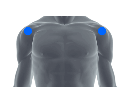
Frozen Shoulder (Adhesive Capsulitis)
The shoulder joint is made up of the head of humerus (ball) and the glenoid fossa (socket). A capsule that holds the ball in the socket. Frozen shoulder is the term used to describe what happens when the shoulder is unable to move due to capsular constriction. Frozen shoulder can be a result of:
» Trauma - either previous surgeries or injuries that did not heal properly
» Overuse
» No specific cause for the pain and stiffness
Symptoms:
» Pain
» Inability to move the shoulder in any and all directions, both passively and actively
Rotator Cuff Tear
There are four muscles that make up the rotator cuff. They are the subscapularis, supraspinatus, infraspinatus and the teres minor. These muscles help to center the ball in the socket as well as work to move and rotate your arm. It is important to know that the primary actions of these muscles are to stabilize the shoulder dynamically. The mechanism of injury for a rotator cuff tear can be the result of falling on your arm, lifting a heavy object, and/or using your shoulder in a repetitive overhead motion. Sports, such as baseball, swimming, and tennis, can also cause rotator cuff injury as well as the grind of everyday activity. A good percentage of rotator cuff injuries do not have a single traumatic event that can cause them.
Symptoms:
» Constant pain in the arm and shoulder which can be worse with certain activity
» Weakness in the arm and shoulder
» Loss of range of motion in your shoulder, especially with overhead motion
Shoulder Impingement
Shoulder impingement occurs when the rotator cuff pinches into the shoulder blade resulting in the pinched rotator cuff becoming inflamed. This inflammation results in pain of the shoulder region. Shoulder impingement can be a result of overhead sports such as baseball, swimming, and tennis. It can also result from activities of daily living such as painting and housework. Aging is also a major contributing factor, as well as someone's bone structure.
Symptoms:
» Minor pain that is present both with activity and at rest
» Pain radiating from the front of the shoulder to the side of the arm
» Sudden pain with lifting and reaching movements
» Pain with overhead sports for e.g. throwing or serving a tennis ball
-
-
Elbow
-
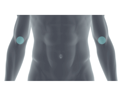
Tennis Elbow
Tennis elbow, also known as lateral epicondylitis, is an inflammation of the wrist extensor tendon which is the muscle that connects to the outer portion of your elbow. The symptoms that result from this inflammation can typically radiate down the forearm from the outside of the elbow. This condition can also cause weakness of the wrist and hand. Tennis elbow can occur due to a repetitive motion activity while one is simultaneously gripping an object. An example would be swinging a tennis racket or using tools such as a hammer.
Symptoms:
» Pain or tenderness on the outside of the elbow
» Pain made worse by lifting a heavy object
» Pain when gripping objects
» Pain that shoots down the outside of the forearm
-
-
Hand & Wrist
-
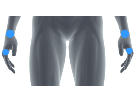
Hand Therapy and Carpal Tunnel Syndrome
Carpal tunnel syndrome is a common disorder affecting the hand. Unlike sprains and tendonitis, which involve injury or inflammation of ligaments or tendons, carpal tunnel syndrome is caused by nerve compression at the wrist.
The carpal tunnel is a small anatomical passageway created by the arch-like arrangement of the small bones that make up your wrist. A tough ligament called the transverse carpal ligament forms the roof of this tunnel. The median nerve passes through this tunnel and can become compressed. This is called carpal tunnel syndrome.
Symptoms:
» Pain, numbness and/or tingling especially in the thumb, index, long and ring fingers. First signs often include waking with pain radiating into the hand from the wrist and/or numbness and tingling in these fingers. This can initially be resolved by shaking or moving the hand around. Symptoms can progress, causing numbness/tingling that is more constant, as well as subsequent hand weakness.
-
-
Hip
-
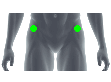
Groin Strain
A strain is the tearing or stretching of muscle fibers. When someone strains the groin muscles, he/she typically is straining the hip adductors (the muscles that bring your legs back toward your midline). The specific muscles that are most commonly affected with a groin strain are the sartorius and the adductor magnus. A groin strain is often described as a pulled muscle by the individual. Groin strains often occur with quick change in direction movements in sports and can also occur with basic running and jumping activities.
Symptoms:
» Pain and tenderness along the inner side of your thigh or groin
» Pain when lifting up the knee or bringing the legs together
» Athletic activity
Bursitis
Bursitis is inflammation of the bursa. The bursa is a fluid filled sac which acts as a cushion between tendons, bones and skin. Bursitis in the hip region is inflammation to the bursa that lies on top of the greater trochanter, a protuberance of the femur. This bursa often becomes irritated due to muscle rubbing against the femur. This injury can occur from running, walking or bicycling.
Symptoms:
» Pain and tenderness on the upper outer area of your thigh
» Increased pain when walking down stairs, walking or bicycling
Hip Arthritis
The hip is a ball and socket joint. In the hip joint, the head of the femur (ball) sits in the acetabulum (socket). The acetabulum or socket has a cartilage lining which allows for the smooth motion of the hip when moving into flexion, extension or abduction. Over time the cartilage becomes damaged and worn down which results in extreme pain that affects everyday living.
Symptoms:
» Chronic, constant and progressive hip pain
-
-
Knee
-
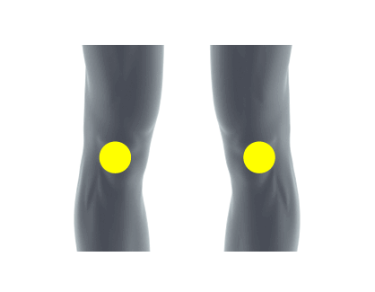
Meniscus Tear
The meniscus is a cartilage block that attaches onto the top of the tibia and makes contact with the femur. There is a lateral and medial meniscus. The meniscus cushions and stabilizes the joint and acts as a shock absorber. As a result, the meniscus reduces the amount of stress on the joint. It is important for joint congruency, load transmission and joint stability. It also serves as an extension and flexion block.
Symptoms:
» Pain which is more common on the medial or inside of the knee
» Swelling within the joint
» Inability to achieve full leg flexion or extension
» Feeling as if your knee gets stuck or locks
» Knee stiffness
» A sensation of buckling or instability in the knee
Patella-Femoral Syndrome
The patella or "knee cap" is held in place by the quadriceps (above) and patellar tendon (below) while sitting in a groove or track on the femur. When patella femoral pain occurs, it is usually the result of mal-tracking of the patella in that very groove in which it sits. Some causes of this mal-tracking can be weak quadriceps, tight IT band and hip/knee structural and anatomical factors.
Symptoms:
» Pain behind or under the knee cap
» Pain while sitting for a long of period of time
» Pain is worse when moving downhill or down stairs
» Popping, snapping, or grinding in the front of the knee
Anterior Cruciate Ligament
The ACL is located in the center of the knee and is a major knee joint stabilizer. The ACL connects the femur to the tibia and checks:
» Anterior translation of the tibia on the femur
» Internal and external rotation of the tibia
» Hyperextension of the knee joint
Most often, the ACL is damaged when the foot is planted and someone or something strikes the knee. Also, a sudden stop or sharp turn with the foot planted can tear the ACL. ACL "non-contact" injuries occur more often in females than in males.
Symptoms:
» Instability in the knee, "giving - out" sensation of the knee
» Pain in the knee
» Swelling within the joint
» Discoloration
Knee Arthritis
The knee has 2 joints, the tibiofemoral joint and the patella-femoral joint. The tibiofemoral joint is where the femur articulates with the tibia. The patella-femoral joint is where the patella articulates with the femur. The cartilage that covers these joints can be damaged and worn down over time and cause chronic pain and impede daily living.
Symptoms:
» Chronic knee pain with activity and at rest
-
-
Foot & Ankle
-
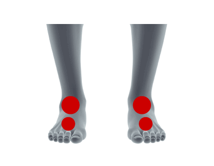
Plantar Fasciitis
The sole of your foot is covered by a fascia. This provides support to the arch of the foot and can become inflamed for various reasons. This inflammation of this fascia is called "plantar fasciitis." It is most commonly inflamed due to overuse or from an increase in the amount of walking, standing and/or stair climbing. Runners are more susceptible to plantar fasciitis due to the amount of stress they put on their feet. This can be a result of a change in running surface, increase in mileage and/or not changing running shoes. Also, aging can insidiously cause this condition, collapsing the arch of the foot, thus putting the fascia on stretch and inflaming it.
Symptoms:
» Pain in the arch of the foot, more significantly in the morning time
» Chronic heel pain
Ankle Sprain
The ankle joint is a complicated joint. There are many ligaments that connect the bones of the foot and ankle together. These ligaments and are very susceptible to injury. The most common mechanism of injury for the ankle joint is an inversion ankle sprain. This means that your ankle is plantar-flexed and inverted (pointed down and in) at the time of injury. Usually, this type of sprain results in a sprain or tearing of the ATF (anterior talofibular ligament) ligament.
Symptoms:
» Pain especially on the outside of the ankle (inversion sprain)
» Swelling
» Discoloration
» Loss of mobility in the ankle joint
Chronic Ankle Laxity
Chronic ankle laxity is instability of the ankle joint. Instability occurs due to previous ankle injuries/sprains resulting in the ligaments of the ankle being stretched and not being able to provide the necessary support to the ankle complex.
Symptoms:
» Feeling of ankle giving way or feeling unstable with various activities
» Continual swelling
» Chronic pain
-